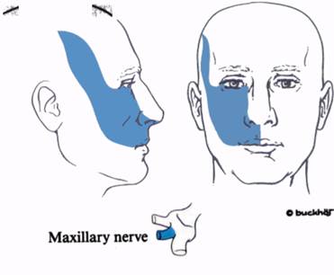
Branches of the maxillary nerve include the posterior, middle and anterior superior alveolar (dental) nerves, which supply the teeth in the upper jaw, together with the adjacent gums and mucous membrane. The anterior superior alveolar nerve also supplies the lateral wall and floor of the nasal cavity, together with part of the septum (Fig. 61:1).
Through its branches to the pterygopalatine ganglion, the maxillary nerve also supplies sensory fibres to the palate (both hard and soft), the tonsil and the lining of the posterior nasal cavity. The main nerves involved are the greater and lesser palatine nerves.
Patient position
Supine.
Landmarks
1. The zygoma
2. The anterior border of the ramus of the mandible, which is easily felt by opening and closing the mouth.
Needle insertion
At the junction of the zygoma and the anterior edge of the ramus, a 7 cm needle is inserted medially while inclined upwards and backwards. At about 4 cm the needle point will contact the sphenoid bone and paraesthesia may be elicited (Fig. 65:1).
Drugs and dose
After aspiration 4 ml of l°7o lidocaine or 0.25% bupivacaine or their equivalent (see p. 20) is injected.
Infraorbital nerve block
Anatomy
The infraorbital nerve runs forward in the infraorbital groove and canal. It enters the face through the infraorbital foramen (Fig. 65:2). It lies under the levator labii superioris and its terminal branches supply the skin of the ala of the nose, the lower eyelid, the cheek and the upper lip. It also supplies the mucous membrane on the inside of the cheek and upper lip.
Landmarks :
1. The lower border of the orbit.
2. The infraorbital foramen.
Needle insertion.
This may be through the skin of the face or through the mouth.
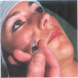
The transcutaneous approach (Fig. 65:4) is made by inserting the needle at the midpoint of the lower border of the orbit, 1 cm below that border. The infraorbital foramen can usually be palpated at this point. The needle is directed upwards to be close to the foramen and paraesthesia may be elicited.
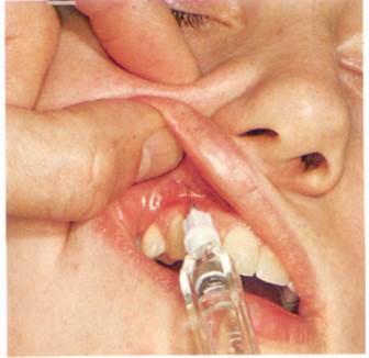
In the transoral approach (Fig. 65:5) the needle is inserted through the superior buccal sulcus and directed upwards to the infraorbital foramen, which is being palpated with the other hand. The needle tip can be felt as it approaches the foramen.
Because of the retrograde spread through the infraorbital foramen the incisor, canine and premolar teeth and the surrounding gums will also be anaesthetised (supplied by the anterior alveolar nerves).
Drugs and dose
After aspiration 2-3 ml of 1% lidocaine or 0.25% bupivacaine or their equivalent (see p. 20) is injected.
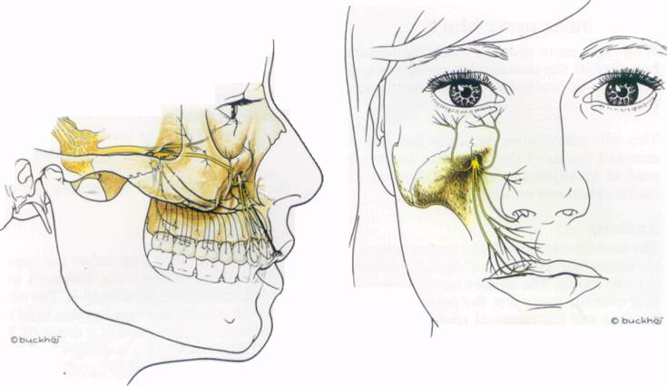
Fig. 65:1
Fie. 65:^
Fig. 65:2.
Pi.. £{••)
Fig. 65:5.
LMMB^fli^M
Mandibular nerve block
While it is more usual to block the terminal branches of the mandibular nerve, the main nerve can be blocked as it leaves the foramen ovale.
This will anaesthetise the lower jaw and the skin and tissues of the lower face and can be used as an adjunct to general anaesthesia in major operations on the jaw.
Anatomy
The mandibular nerve is the largest branch of the trigeminal nerve (Fig. 67:1). It is both sensory and motor. The sensory nerves supply the skin over the lower jaw, the posterior part of the face and the temporal region, (Fig. 67:2) the mucous membrane of the lower lip and the floor of the mouth, and the lower teeth and gums. The motor branches supply the muscles of mastication.
The large sensory root leaves the cranium through the foramen ovale, where it is joined by the motor root. After giving off a meni ngeal branch and the nerve to the medial pterygoid muscle, the mandibular nerve divides into an anterior and a posterior trunk. The sensory fibres of the anterior trunk run in the buccal nerve, which is distributed with branches of the facial nerve. It supplies the skin and mucous membrane on either side of the buccinator muscle and the posterior part of the buccal surface of the gum of the lower jaw. The motor fibres supply the masseter and temporalis muscles.
Уважаемый посетитель!
Чтобы распечатать файл, скачайте его (в формате Word).
Ссылка на скачивание - внизу страницы.