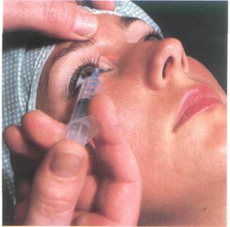
The needle (23 gauge, 4 cm) is inserted perpen-dicularjy backwards at the upper and medial part of the orbit, between the eyebrow and the palpebral fissure, closer to the former than the latter (Fig. 61:2). At a depth of about 1.5 cm the bone of the orbit will be contacted. If the bone is not contacted at this depth, the needle should be withdrawn and directed more medially until the medial wall of the orbit is felt at the depth of 1.5-2 cm. The optic nerve is 4 cm deep to the palpebral fissure.
Drugs and dose
After aspiration inject 2 ml of 1% lidocaine or 0.25% bupivacaine or their equivalent. (See p. 20).
Fig. 61:1. Courtesy of Astra
1. Supraorbital nerve
2. Frontal nerve
3. Lacrimal nerve
4. Nasociliary nerve
5. Maxillary nerve
6. Zygomatic nerve
7. Infraorbital nerve
8. Lateral branch of the frontafherve
9. Medial branch of the frontal nerve
10. Supratrochlear nerve
11. Infratrochlear nerve
12. Nasopalatine nerve
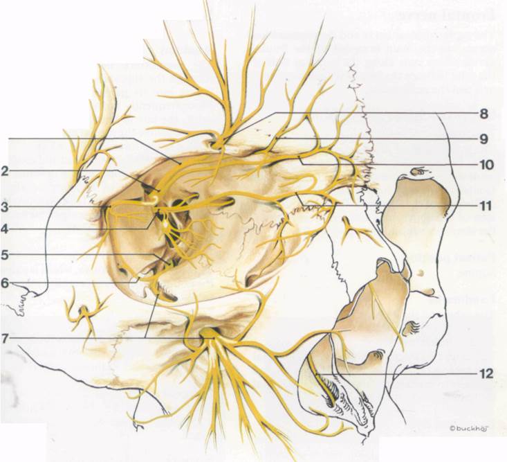
Fig. 61:1.
Fie. 61:2.
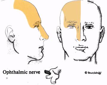
Frontal nerve
The supratrochlear nerve and the supraorbital nerves are the main branches of the frontal nerve, which runs along the superior wall of the orbit between the levator palpebrae superiT oris and the periosteum.
Supratrochlear nerve block
Anatomy
The supratrochlear nerve emerges from the orbit at its upper and medial part and runs upward under the frontal belly of the occipito-frontalis muscle to supply the skin of the medial aspect of the forehead. It also supplies the skin of the upper nose and sends branches to the conjunctivae and the upper eyelid.
Patient position
Supine.
Landmarks
The orbit and the eyebrow.
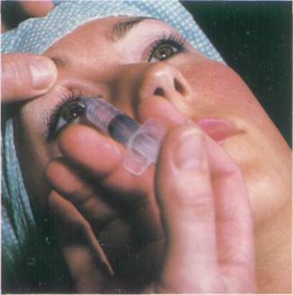
Needle insertion
The needle is inserted at the upper and medial part of the orbit. It is directed upwards and medially to contact the frontal bone just lateral to the root of the nose (Fig. 63:2).
Drugs and dose
After aspiration 2-3 ml of 1% lidocaine or 0.25% bupivacaine or their equivalent (see p. 20) is injected.
Supraorbital nerve block
Anatomy
The supraorbital nerve can leave the orbit through the supraorbital foramen before dividing into its medial and lateral branches. More frequently it divides before leaving the orbit, the lateral branch leaving through the foramen while the medial branch emerges 1 cm medial to the foramen (Fig. 63:1).
It supplies the upper eyelid and conjunctiva, before dividing and running upwards deep to the frontal belly of occipitofrontalis. After reaching the superficial fascia the two branches supply the skin of the scalp as far back as the lambdoid suture (Fig. 63:4).
Landmarks
The supraorbital foramen, which is palpable at the midpoint of the supraorbital margin of the orbit.
Needle insertion
While palpating the foramen the needle is inserted just under the eyebrow and directed upwards to lie close to the foramen (Figs. 63:3).
Drugs and dose
After aspiration 2-3 ml of 1% lidocaine or 0.25% bupivacaine or their equivalent is injected.
Superficial block of the supratrochlear and supraorbital nerves
The terminal cutaneous branches of these nerves, which supply the forehead and scalp, become superficial just above the eyebrow. They may be blocked by a subcutaneous infiltration in a horizontal line, 2 cm above the eyebrow from the lateral border of the orbit to the midline (Fig. 63:5).
Fig. 63:1. Courtesy of Astra.
1. Lateral branch of the supraorbital nerve
2. Medial branch of the supraorbital nerve
3. Supratrochlear nerve
Fig. 63:4. Courtesy of Astra.
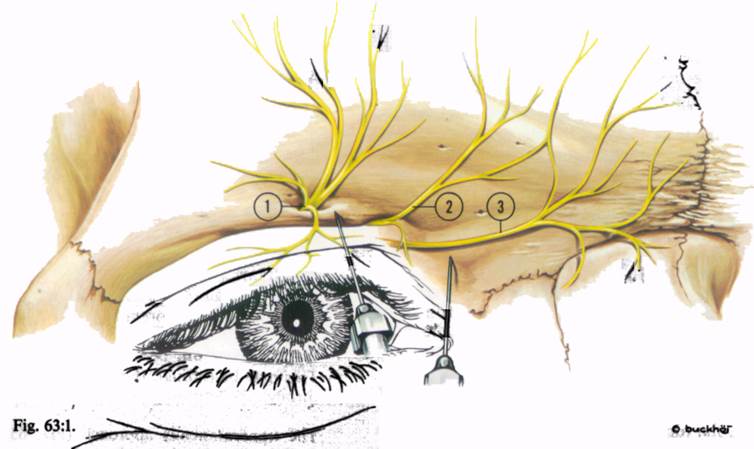
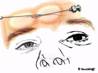
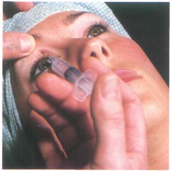
Fig. 63:2. Fig. 63:4.
Fig. 63:3. Fig. 63:5.
Maxillary nerve block
While it is possible to block the terminal branches of the maxillary nerve, (i.e. the infra-orbital, superior alveolar and palatine nerves), the maxillary nerve itself can be blocked more simply as it crosses the pterygopalatine fossa. This anaesthetises both the skin and the deep structures of the middle face, (Fig. 65:3) including the nasal cavity, the maxillary bone and sinus, the upper teeth, and the upper part of the mouth and the oral cavity.
Anatomy
The maxillary nerve leaves the cranium through the foramen rotundum, crosses the pterygopalatine fossa and enters the orbit through the inferior orbital fissure (Fig. 57:1), where it becomes the infraorbital nerve.
Уважаемый посетитель!
Чтобы распечатать файл, скачайте его (в формате Word).
Ссылка на скачивание - внизу страницы.