To block all three nerves with a single injection, the needle must reach the trigeminal ganglion by passing through the foramen ovale. Using a nerve stimulator, it is possible to identify the three main branches of the trigeminal nerve.
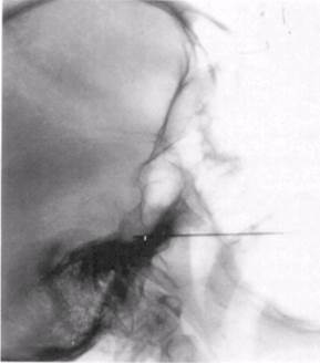
Because of the accuracy required in locating the foramen ovale, and the serious consequences of a misplaced injection, this block should only be done under radiographic control.
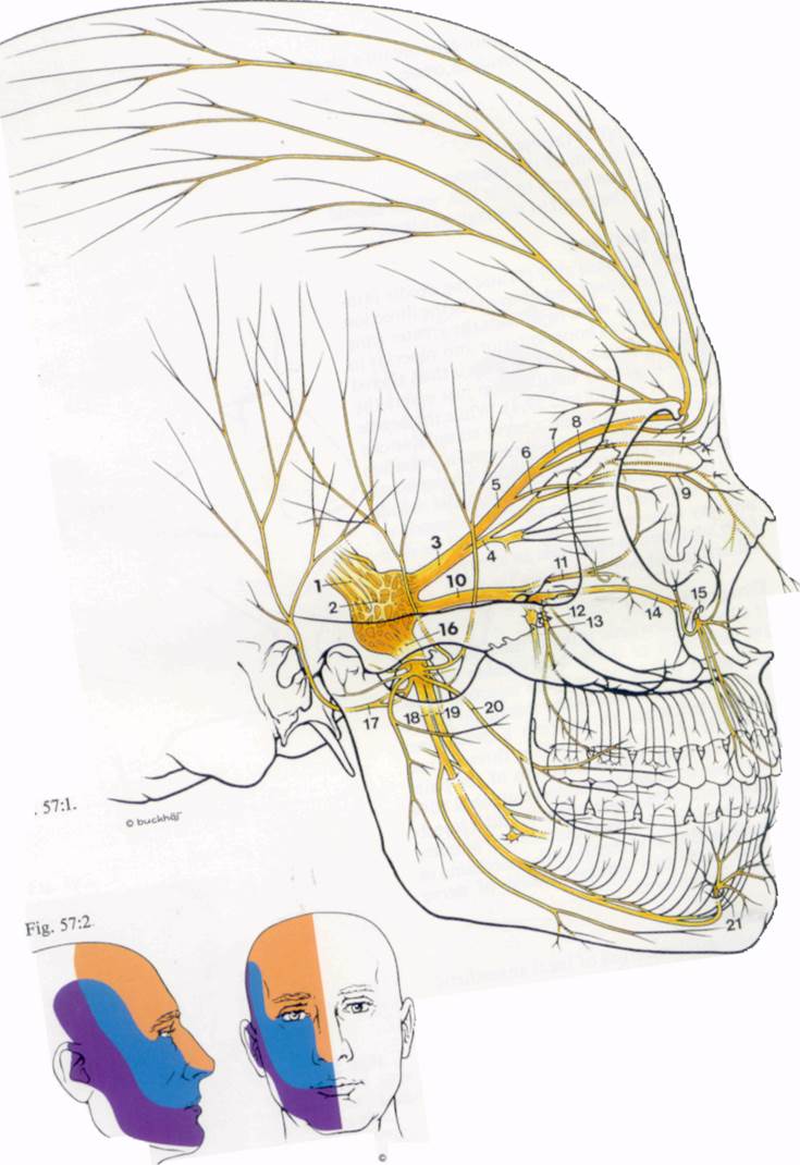
Fig. 57:1.
1. Trigeminal nerve
2. Trigeminal (Gasserian) ganglion
3. Ophthalmic nerve
4. Nasociliary nerve
5. Supraorbital nerve
6. Lacrimal nerve
7. Frontal nerve
8. Supratrochlear nerve
9. Infratrochlear nerve
10. Maxillary nerve
11. Zygomatic nerve
12. Middle superior alveolar nerve
13. Posterior superior alveolar nerve
14. Anterior superior alveolar nerve
15. Infraorbital nerve
16. Mandibular nerve
17. Auriculotemporal nerve
18. Inferior alveolar nerve
19. Lingual nerve
20. Buccal nerve
21. Mental nerve
Fig
b^cW^i
SSS^
Patient position
Supine with head in position to obtain a clear X-ray picture of the foramen ovale.
Landmarks
Fig. 59:3. i
1. The pupil of the eye.
2. The midpoint of the zygoma.
3. Point 3 cm lateral to the lips, which should level withlhe pupil in the sagittal plane.
Needle insertion
A wheal is raised over (3) and the needle is inserted backwards and upwards in the direction of (1) and (2), so as to contact the greater wing of the sphenoid bone, anterior and superior to the foramen ovale. The direction is then altered using radiography until the tip just enters the foramen (Fig. 59:2 and 59:4). While the needle may elicit paraesthesia, a nerve stimulator can be used to identify the three main branches leaving the Gasserian ganglion. The ophthalmic branch is superior and medial, the mandi-bular is inferior and lateral, with the maxillary midway between. The needle should be carefully aspirated to eliminate an intravascular or subarachnoid position of the tip.
Drugs and dose
If a local anaesthetic is being used, 0.5-1 ml of 2% lidocaine or 0.5% bupivacaine or their equivalent (see p. 20) may be injected. However, it is more usual in the treatment of tri-geminal neuralgia to perform a permanent neurolysis of one or more of the three nerves, depending upon the distribution of the pain. The choice of agent lies between alcohol 80% and phenol 5-10% in glycerine. 0.1 ml is injected and the effect assessed over several minutes before injecting a further 0.1 ml, continuing in this way until the required extent of nerve blockade is achieved.
Complications
1. Subarachnoid injection of local anaesthetic at the base of the brain can lead to 'unconsciousness and blockade of the ipsilateral cranial nerves.
2. Blockade of the ophthalmic nerve renders the eye analgesic and can lead to corneal ul-ceration.
Figs. 59:3.
/. The pupil of the eye.
2. The mid point of the zygoma.
3. Point 3 cm lateral to the lips, which should be level with (1) in the sagittal plane.
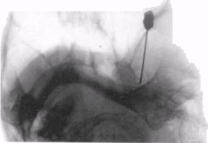
Fig. 59:2. Courtesy of Dr. G.L.M. Carmichael Fig. 59:4. Courtesy of Dr. G.L.M. Carmichael
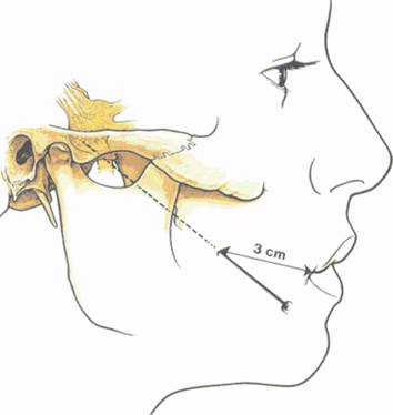
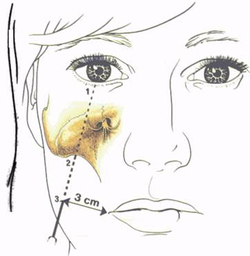
Fig 59:1
Fig.
Fig 59:2
59:4
Ophthalmic nerve
Although the ophthalmic division of the tri-geminal nerve can be permanently blocked close to the trigeminal ganglion, there are few indications.for such a procedure, given the dangers of anaesthetising the eye with the possibility of corneal ulceration.
The branches of the ophthalmic nerve, however, which supply the skin of the upper eyelid, the forehead and the side of the nose, are readily blocked and this is of use in plastic surgery. The main branches are the nasociliary and the frontal nerves (Fig. 61:1).
Nasociliary nerve
The nasociliary nerve supplies the skin and mucous membrane of the nose, the cornea and the conjunctiva. Two internal nasal branches supply the mucous membrane of the anterior part of the nose, including the nasal septum. An external nasal branch supplies the skin of the lower part of the nose below the nasal bone. The long ciliary nerves supply the cornea and conjunctiva.
Patient position
Supine.
Landmarks
The orbit, the eyebrow and the medial palpe-bral fissure.
Needle insertion
Уважаемый посетитель!
Чтобы распечатать файл, скачайте его (в формате Word).
Ссылка на скачивание - внизу страницы.