GLASS STRENGTHENING WITH AN ULTRAFAST LASER
Paper M404
Science & Technology, Corning Incorporated, Corning, NY 14831, USA
Abstract
We performed laser-strengthening experiments on a number of glasses and glass-ceramics. The objective of this study was to identify the mechanism of strengthening and to establish the correlation between the residual stress and the apparent fracture-toughness increase. After the laser exposure, the stress was measured by interpreting the resulting birefringence pattern (Polscope™). The apparent fracture toughness was measured by a Double-Torsion method, which required a minimal number of samples to reliably detect the effect of laser treatment as compared with the four-point bending test. We discovered that the apparent fracture-toughness increase is proportional to the magnitude of laser induced stress. We also present the results on how the glass strengthening is influenced by the irradiation pattern and laser parameters.
Introduction
In 2005, a group from Japan presented the results of glass strengthening using an ultrafast laser [1]. A sodalime thin glass sample was irradiated with the focused beam of a femtosecond laser and glass structure was altered in the focal area. In the four-point bending test the authors observed ~ 50% increase in the static fracture toughness, which they attributed to crack arrest by these densely spaced spots of modified glass. We expanded experiments to a number of glasses and glass-ceramics (GC) in order to further explore this effect, to understand the mechanism of strengthening, and to study the strengthening response of different glasses and glass-ceramics to the femtosecond-laser exposure.
Experimental
For laser treatment, we utilized the amplified 800 nm femtosecond laser system, which operated at 20 kHz repetition rate with variable pulse energy of up to 25 µJ. The pulse duration was 40-50 fs. The samples were placed on the Aerotech motorized XYZ translation stage using a vacuum chuck. The laser beam was focused onto the sample with the NA=0.26 objective.
Most of the experiments were carried out on the 1-mm thick microscope slides made of 0215 soda-lime glass. This choice was dictated by the availability of the samples and because this glass is well characterized. We also explored 0317 soda aluminosilicate glass, 9664 Spinel glass precursor, and 9664 Spinel GC. The thickness of all of them was about 1 mm. The size of the samples was 1” by 3” and the reason for this choice is discussed below. All these materials are transparent at the laser wavelength and the structural modification was introduced through nonlinear absorption of intense ultrashort optical pulses [2]. The materials that we studied are listed in Table 1 below.
Table 1. Glasses and glass-ceramics used in experiments.
|
Code 0215 |
Code 0317 |
9664 glass |
9664 GC |
|
|
Density, g/cm3 |
2.4 |
2.53 |
2.69 |
2.75 |
|
CTE, x10-7 ºC-1 |
89 |
88 |
~26 |
37 |
|
Strain point, ºC |
511 |
508 |
734 |
920 |
|
Annealing point, ºC |
545 |
550 |
781 |
>1000 |
|
Softening point, ºC |
724 |
720 |
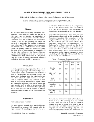
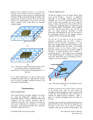
Two exposure patterns were produced in glasses as shown in Fig.1. The scanning lines are oriented differently in the two cases. In the first case (parallel), the lines are parallel to the surface of the plate (Fig. 1 (a)) while in the second (perpendicular) case the lines are perpendicular to the surface (Fig. 1 (b)). The laser beam was propagating in the negative Z direction. The patterns were covering the area of 3.5 x 3.5 mm. The thickness of the structures and their alignment were such that the laser-written structures were buried inside the glass and did not protrude through the surface. The gap between the patterns and the front and the back surfaces was approximately 20 µm and it prevented surface damage, which could affect the measured strength of glass.
(b)
Fig. 1. The exposure patterns produced in glasses with the femtosecond laser: (a) parallel and (b)
perpendicular. The laser beam is propagating in the –Z direction.
In our initial experiments, we did not observe any strengthening effect from the “perpendicular” pattern and we used the “parallel” irradiation pattern in all experiments described below.
Characterization
Stress measurements in the glass samples were carried out using the PolScope™ technique [3]. The microscopic images are taken at different polarization conditions and the software calculates map of retardation and of slow-axis orientation. Knowing the stress-optic coefficient, the retardation is converted into stress. In all studied materials, laser treatment resulted in compressive stress.
Уважаемый посетитель!
Чтобы распечатать файл, скачайте его (в формате Word).
Ссылка на скачивание - внизу страницы.