|
Anatomy |
Анатомия |
|
The cells of the body must receive a continuous supply of oxygen and nutrients. Waste products must be removed, also. It is the circulatory system that transports the blood from the lungs to all parts of the body with its life giving oxygen. The average adult contains over 60,000 miles of blood vessels. The pump that propels the blood through this long network of vessels is the heart. |
Сердце – основной орган системы кровообращения. Оно выполняет насосную функцию, обеспечивая циркуляцию крови по сосудам, общая протяженность которых составляет свыше 35 000 км. |
|
Heart |
Сердце |
|
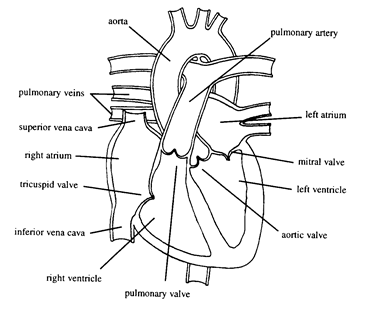
The heart is cone shaped and found between the lungs in the middle of the chest, behind the sternum, in the middle mediastinum. The heart lies with the blunt point, the apex, pointing downward and to the left. Approximately, two thirds of its mass is in the left thoracic cavity. The heart of the average male weighs about 280-340 grams. The heart of the average female weighs about 230-280 grams. The dimensions of the heart are a length of 12 cm, a width of 8 cm and a thickness of 6 cm. There are four chambers in the heart with four one-way valves to direct blood flow through the chambers. The upper chambers are the right and left atria. They are divided by a thin muscular wall. the atrial septum. The septum contains a central depression of thin fibrinious material known as the fossa ovalis. |
Сердце расположено в переднем средостении. Масса сердца взрослого мужчины в среднем 280-340 г, взрослой женщины – 230-280 г. Примерно 2/3 массы сердца составляет миокард левого желудочка. Сердце имеет четыре камеры. Кровь поступает в предсердия, затем в желудочки. Левое и правое предсердия разделены межпредсердной перегородкой. В средней части перегородки имеется участок овальной формы, состоящий из соединительной ткани - fossa ovalis. |
|
The lower chambers are the right and left ventricles. They are divided by the ventricular septum. This is a thick muscular wall with a small, thin membranous portion at the top. |
Правый и левый желудочек разделены межжелудочковой перегородкой. Межжелудочковая перегородка имеет достаточную толщину, состоит из мышечной ткани. В верхнем отделе ее имеется небольшой участок, представленный тонкой соединительнотканной мембраной. |
|
Frontal View |
Передняя проекция |
|
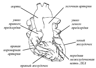
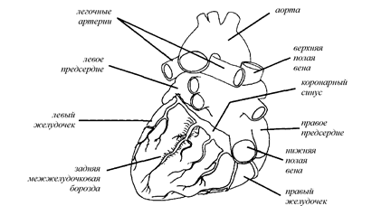
Most heart surgery techniques require opening the sternum to expose the heart. This surgical opening, the median sternotomy displays the right atrium and right ventricle. Seen in the drawing below are the right atrium and right ventricle separated by the right atrioventricular sulcus. This sulcus is a groove in which the right coronary artery runs. Further left is the anterior interventricular sulcus separating the right and left ventricles. The anterior descending branch of the left coronary artery runs through this sulcus. The left atrium and ventricle have only small portions visible in this view because they lie posterior to the right atrium and ventricle. |
При большинстве сердечно-сосудистых операций применяют срединную стернотомию, дающую доступ к правым отделам сердца (рис. 1). На рисунке виден правый желудочек и правое предсердие, разделенные правой предсердно-желудочковой бороздой, в которой проходит правая коронарная артерия. Правый и левый желудочек разделены межжелудочковой бороздой, в которой проходит передняя нисходящая ветвь левой коронарной артерии. В данной проекции видна лишь небольшая часть левого предсердия и левого желудочка. |
Рисунок 1. Сердце – вид спереди
|
|
|
Posterior View |
Задняя проекция |
|
In the posterior view below, large portions of the left atrium and ventricle are visible. Also seen are the pulmonary veins, the coronary sinus lying in the posterior atnoventncular sulcus. and the posterior interventricular sulcus containing the descending branch of the right coronary artery. |
В задней проекции (рис. 2) видны левые отделы сердца – левое предсердие и левый желудочек, а так же легочные вены и коронарный синус, расположенный в задней предсердно-желудочковой борозде. Нисходящая ветвь правой коронарной артерии располагается в задней межжелудочковой борозде. |
|
Posterior View |
Рисунок 2. Сердце – вид сзади |
|
|
|
|
Heart Walls |
Строение сердца |
|
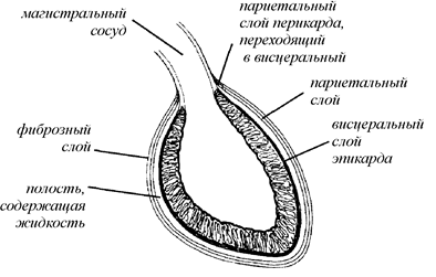
The heart has a protective covering, the pericardium. This two layered covering allows the heart to move with a minimum of friction. The loose outer layer, the fibrous layer, is a tough white connective tissue. This layer becomes continuous with the outer layers of the great vessels. |
Сердце находится в полости перикарда – оболочки из соединительной ткани, состоящей из наружного и внутреннего слоя. Наружный слой состоит из плотной белой соединительной ткани, он продолжается по ходу крупных сосудов. |
|
The inner layer is the serous pericardium. This layer is further divided into the parietal and visceral layers. The parietal layer covers the fibrous layer. It runs superiorly under the fibrous pericardium for approximately 2 cm up the great vessels, then turns back upon itself. At this point it now becomes the visceral layer or epicardium, and continues back to cover the heart. Both layers of the serous pericardium are moist, smooth and mesothelial covered. Between the parietal layer and the epicardium is a space containing approximately 50 ml of pericardial fluid. |
Внутренний слой представляет собой серозную оболочку, имеющую, в свою очередь, два слоя. Париетальный слой прилежит к фиброзному слою перикарда, простирается примерно на 2 см по ходу крупных сосудов, затем перходит в висцеральный слой – эпикард, покрывающий сердце снаружи. Париетальный и висцеральный слой перикарда покрыты мезотелием и имеют гладкую поверхность. Полость между ними заполнена жидкостью, количество которой составляет около 50 мл. |
|
There are three layers of the heart wall. The outer most layer is the epicardium (the visceral pericardium). This is a single layer of cells of mesothelial cells over a fibroelastic membrane. The cardiac muscle wall is the myocardium. This muscle is similar to the voluntary muscles. The myocardium muscle cells are elongated and interconnected to allow coordinated contractions of the heart. There are two bands of muscles that form a latticework arrangement to accomplish these contractions. The inner most layer is the endocardium. This is a single layer ofendothelial cells that creates a very smooth lining of the interior chambers of the heart. This smooth lining allows blood to move through the heart with a minimum of friction. Lesions of the endocardium caused by inflammation can cause local thrombosis and thromboembolism. |
Стенки сердца состоят из трех слоев. Наружный слой – эпикард (висцеральный перикард) – представляет собой мезотелий. Основной слой – миокард, он представляет собой синцитий из поперечнополосатых мышечных волокон. Изнутри полости сердца выстилает эндокард – слой эндотелиальных клеток. При воспалении эндокарда на нем образуются тромбы, вызывающие тромбоэмболию. |
|
Рисунок 3. Строение сердца (схема)
|
|
|
Right Heart |
Правые отделы сердца |
|
The superior vena cava returns blood from the upper body to the right atrium. Blood from the head, arms and upper body is drained into the superior vena cava. The inferior vena cava drains blood from the lower body into the right atrium. The right atrium is the small chamber that is the site of venous cannulation for CPB. The walls of the right atrium are thin, about 2-3 millimeters. |
В правое предсердие впадают верхняя и нижняя полые вены. Верхняя полая вена дренирует верхнюю часть туловища, голову, верхние конечности. Нижняя полая вена дренирует нижнюю часть туловища, нижние конечности. Правое предсердие имеет тонкие стенки (2-3 мм) и относительно малый размер полости. Через правое предсердие канюлируют полые вены. |
|
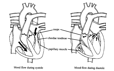
The blood moves from the right atrium through an atrioventricular (A-V) valve. A-V valves have leaflets which tethered by fibrous chords, the chordae tendineae that are attached to the papillary muscles in the walls of the ventricle. These chordae tendineae and papillary muscles prevent the leaflets from going back into the atrium when the heart contracts. Thus, a complete closure occurs. This first A-V valve is the tricuspid valve which has 3 leaflets as its name implies. |
Полости правого предсердия и правого желудочка сообщаются через правое атриовентрикулярное отвестие, закрытое в диастолу трикуспидальным клапаном. Створки клапана с помощью сухожильных хорд соединены с папиллярными мышцами, исходящими из стенки желудочка. Сокращения папиллятных мышц удерживают в систолу створки клапана в закрытом состоянии. |
|
Atrioventricular Valve |
|
|
|
|
|
The right ventricle is the lower chamber of the right heart. The top part of the ventricle surface is smooth, but in the lower portion bundles of muscles arise from the walls to form interlocking webs. Contraction of the right ventricle forces blood through the pulmonary valve. |
Во время систолы кровь из правого желудочка поступает в легочную артерию. Верхние отделы правого желудочка имеют гладкую внутреннюю поверхность, нижние – трабекулярное строение. |
|
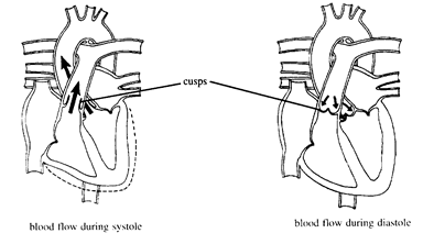
A semilunar valve, the pulmonary valve, allows blood to enter the pulmonary artery. This valve has three cusps that form pockets. These cusps balloon out as they are filled and come together to prevent backflow into the ventricle at the end of systole. These half-moon shaped cusps give this type of valve its name, semilunar valve. The pulmonary artery transports blood from the right ventricle to the lungs. This is the only artery that transports venous blood. This is a short vessel, only about 2 inches long. It branches into the right and left pulmonary arteries. These enter their respective lung near the base. |
Клапан легочной артерии (полулунный) нахолится в устье легочной артерии. Он состоит из трех створок, напоминающих карманы, препятствующие ретроградному току крови. Венозная кровь по легочной артерии поступает в легкие. Ствол легочной артерии относительно короткий, около 5 см у взрослых, и делится на левую и правую легочную артерию. |
|
Semilunar Valve |
|
|
|
|
|
Left Heart |
Левые отделы сердца |
|
Blood from the lungs that has been oxygenated enters the left atrium through four pulmonary veins. The left atrium is slightly larger than the right atrium During ventricular diastole, when the heart relaxes, blood flows through the mitral valve, an atrioventricular valve, into the left ventricle. This valve has two leaflets held by fibrous chordae tendineae. It is sometimes referred to as the bicuspid valve. The chordae tendineae are attached to the anterior and posterior papillary muscles. The papillary muscles and chordae tendinae assist in ventricular contraction by allowing longitudinal shortening of the left ventricle during systole. Because of this, when a patient has a mitral valve replaced, the posterior leaflet is often spared. This chordal sparing procedure has been shown to help maintain ventricular function. The left ventricle is the most powerful chamber with a wall much thicker than the right ventricle. This ventricle has an extra layer of muscle, the deep bulbo layer, that goes from the aortic and mitral valve rings around the left ventricle. The left ventricle contracts and blood is forced through the aortic semilunar valve into the aorta. |
Оксигенированная в легких кровь поступает по легочным венам в левое предсердие. Полость левого предсердия несколько больше, чем правого. В диастолу через открытый митральный клапан кровь поступает из левого предсердия в левый желудочек. Митральный клапан имеет две створки, от которых отходят сухожильные хорды, крепящиеся к папиллярным мышцам. В систолу папиллярные мышцы способствуют сокращению левого желудочка по его длинной оси, поэтому при протезировании митрального клапана его заднюю створку часто оставляют с целью сохранения сократительной функции левого желудочка. Левый желудочек – самая мощная камера сердца, его стенки имеют наибольшую толщину. В левом желудочке имеется дополнительный глубокий мышечный слой, берущий начало от кольца аортального и митрального клапана. В систолу кровь из левого желудочка поступает в аорту через открытый аортальный клапан, имеющий три створки. |
|
The aortic valve, a semilunar valve, has three cusps that balloon and seal to prevent backward flow. This prevents blood in the aorta from returning to the left ventricle during diastole. The aortic and the pulmonic valves are located in an enlarged portion of their great vessels known as the aortic and pulmonary sinus ofValsalva. |
|
The Heart |
|
|
|
|
|
Coronary Arteries |
Коронарные артерии |
|
The heart, itself, also needs oxygenated blood supplied to its cells. The blood that passes through the chambers of the heart cannot supply the myocardium. The coronary arteries must supply these needs. The coronary arteries come from the aorta. The openings to the coronary arteries are located in the aortic sinus of Valsalva. These openings are called ostia and are found behind the aortic valve cusps. When the valve is closed during diastole, blood flows into the openings to perfuse the coronary arteries. The left and right coronary arteries branch into other arteries and send branches into the myocardium to supply this need. |
Сердечная мышца кровоснабжается по коронарным артериям. Коронарные артерии берут начало в синусе Вальсальвы, их устья находятся позади створок аортального клапана. Кровь в устья коронарных артерий поступает в диастолу при закрытом аортальном клапане. От аорты отходят две основных коронарных артерии – левая и правая, которые, в свою очередь, имеют несколько ветвей, кровоснабжающих различные участки миокарда. |
|
The left main coronary artery supplies the myocardium of the left ventricle. This artery divides into the anterior descending branch and the circumflex branch. The left anterior descending branch runs on the front of the heart, down to the apex. This supplies the apex, anterior surface and anterior septum. The circumflex branch lies in the groove between the left atrium and left ventricle. This branch furnishes a portion of the left ventricle away from the septum with oxygenated blood. |
Левая коронарная артерия кровоснабжает левые отделы сердца. Она имеет две основные ветви – переднюю нисходящую артерию и огибающую артерию. Передняя нисходящая артерия проходит в межжелудочковой борозде по передней поверхности сердца к его верхушке, кровоснабжает переднюю поверхность левого желудочка, верхушку и переднюю часть межжелудочковой перегородки. Огибающая артерия располагается по задней поверхности сердца в левой предсердно-желудочковой борозде, кровоснабжает заднюю поверхность левого желудочка |
|
The right coronary artery is located in the groove between the right atrium and right ventricle. It runs down and around to the back of the heart. The ;come off the right coronary artery and branch to supply the right ventricle. In most cases the right coronary artery terminates in the posterior interventricular groove as the posterior descending artery which supplies blood to the posterior septum. It also may continue and provide branches to the posterior left ventricular wall. These are known as posterior lateral muscular branches. |
Правая коронарная артерия проходит в правой предсердно-желудочковой борозде к задней поверхности сердца, в проксимальном отделе отдает ветви, кровоснабжающие правое предсердие и желудочек. В большинстве случаев правая коронарная артерия заканчивается задней нисходящей артерией, кровоснабжающей заднюю часть межжелудочковой перегородки. Артерия может продолжаться, отдавая заднебоковые ветви, кровоснабжающие заднюю часть левого желудочка. |
|
Coronary Veins |
Коронарные вены |
|
The venous drainage of the heart consists of the coronary sinus system and the anterior cardiac system. The coronary sinus is a large vessel that allows blood from the superficial veins to enter the right atrium. The coronary veins mirror the coronary arteries to some extent. The great cardiac vein runs beside the LAD and drains into the coronary sinus. Also draining into the coronary sinus are the left marginal, left posterior and middle veins. Draining into the anterior cardiac system are the right marginal, small cardiac and sinus node veins. The small cardiac vein runs along the RCA. The cardiac veins do not have valves as many of the other veins of the body have. This makes it possible to give retrograde cardioplegia. |
Венозная система сердца состоит из системы коронарного синуса и переднего бассейна. Коронарный синус – крупный сосуд, собирающий венозную кровь из поверхностных вен и впадающий в правое предсердие. Расположение коронарных вен в некоторой степени повторяет расположение коронарных артерий. Вены сердца не имеют клапанов, что позволяет вводить кардиоплегический раствор через вены ретроградно. |
|
Arteries |
Строение артерий |
|
Arteries start this highway that delivers oxygenated blood. They continue to branch into smaller branches. The arterioles are the smallest branches of the arteries. Capillaries link arterioles and venules. the smallest veins. These microscopic vessels are the smallest blood vessels. |
Крупные коронарные артерии и их ветви заканчиваются множеством мелких ветвей. Мельчайшие артерии – артериолы – оканчиваются сетью капилляров. |
|
Arteries contain 3 layers of tissue. The inside layer, the intima, is a smooth endothelial lining, connective tissue and some muscle. The middle layer, the media, is made of muscle and elastic fibers. This allows expansion of the vessel. The outside layer, the adventitia, is fibrous connective tissue that prevents the vessel from expanding too much. This ability to expand and contract helps regulate blood flow through the body. |
Стенка артерии имеет три слоя. Внутренний слой (интима) состоит из эндотелия, соединительной ткани и небольшого количества мышечных волокон. Средний слой (медиа) содержит мышечные и эластические волокна. Наружный слой (адвентиция) представляет собой соединительную ткань. |
|
Veins |
Строение вен |
|
The blood, after giving up its oxygen and nutrients, picks up carbon dioxide and waste products before returning to the heart. This return trip is by way of the venules that become the larger veins. The vein is thin walled, but it does not contain the high pressures that the arteries must accommodate. Most veins contain valves that prevent backflow of the blood. |
Проходя по капиллярам, кровь доставляет клеткам кислород, эвакуирует углекислый газ и шлаки, затем поступает в венулы и вены. Вены представляют собой тонкостенные сосуды, давление в которых значительно ниже, чем в артериях. Большинство вен (но не вены сердца) содержат клапаны, препятствующие ретроградному току крови. |
|
Aortic Arch Vessels |
Артерии, отходящие от дуги аорты |
|
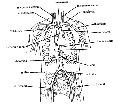
The blood leaves the left ventricle and enters the aorta, the largest artery of the body. This large vessel has a diameter of about 2.5 cm. The portions of the aorta are referred to as the ascending aorta, aortic arch and descending aorta. The descending aorta is further divided into the thoracic aorta and abdominal aorta. The aorta leaves the heart and turns downward. This turn of the aorta is known as the aortic arch. Three arteries branch from the aortic arch. The innominate artery is the first vessel to come off. It divides into the right carotid and the right subclavian arteries. The next artery to come off the aorta is the left common carotid artery. The third artery is the left subclavian artery. The left internal mammary artery branches from the left subclavian artery. |
Аорта взрослого человека имеет диаметр около 2.5 см. Различают восходящую часть аорты, дугу аорты и нисходящую часть аорты – грудную и брюшную аорту. От дуги аорты отходят три крупных сосуда: плечеголовной ствол, которая делится на правую общую сонную артерию и правую подключичную артерию; левая общая сонная артерия; левая подключичная артерия. От левой подключичиной артерии отходит левая внутренняя грудная артерия, которую часто используют в качестве аутоартериального шунта при операции коронарного шунтирования. |
|
The carotid arteries supply much of the blood for the brain. The left and right common carotid arteries branch into external and internal carotid arteries. It is the internal carotid artery that supplies the brain and eyes. The middle cerebral artery branches from the internal carotid. This artery branches to supply the brain. Embolisms causing strokes often occur in these arteries. Branching from the internal carotid artery is the posterior communicating artery. Branches of this artery supply the optic cortex. The external carotid artery supplies the neck, face and exterior of the head. This artery branches into the temporal and internal maxillary arteries. |
Сонные артерии – основные артерии, кровоснабжающие головной мозг. Правая и левая общие сонные артерия делятся на наружную и внутреннюю сонную артерию. Внутренние сонные артерии кровоснабжают головной мозг и глаза. От внутренней сонной артерии отходит средняя мозговая артерия, и задняя коммуникантная артерия, ветви которой кровоснабжают зрительную кору. Наружная сонная артерия кровоснабжает шею, лицо, наружную поверхность головы. От нее отходят височная и внутренняя нижнечелюстная артерии. |
|
Arteries of the Upper Extremities |
Артерии верхних конечностей |
|
The subclavian artery supplies the upper extremity. The subclavian artery becomes the axillary artery at the lateral edge of the first rib. The axillary artery becomes the brachial artery in the upper arm at the tendon of the teres major muscle. Also branching from the axillary artery is the thoracic artery which supplies the chest wall. The radial and ulnar arteries come off the brachial artery just below the elbow. The radial artery runs down the arm and branches into the palmer arch to supply the thumb. The ulnar artery supplies the arm and palm. From the ulnar artery branches the palmar arch of the hand. This arch has two branches. These are the superficial and deep branches. From the palmar arch four digital arteries come off. Each of these arteries extends into a finger. |
Верхние конечности кровоснабжают подключичные артерии. Латеральнее края I ребра артерия называется подмышечной. Здесь она отдает грудную артерию, кровоснабжающую стенку грудной клетки. Продолжение подмышечной артерии – плечевая артерия, которая после локтевого сустава делится на лучевую и локтевую артерию. |
|
Branches From the Descending Aorta |
Ветви нисходящей аорты |
|
The descending aorta is divided into two sections. The first section is the thoracic aorta. The abdominal aorta begins at the diaphragm. The abdominal aorta gives off branches that include the celiac, mesenteric and renal arteries. The celiac artery comes off the aorta near the diaphragm and branches into the gastric, hepatic and splenic arteries. The gastric artery supplies the stomach. The hepatic artery is a large artery that branches into left and right branches to supply the liver. The splenic artery supplies the spleen. The superior mesenteric artery supplies the intestines, while the inferior mesenteric artery furnishes blood to the colon and the rectum. The renal arteries come off the abdominal aorta to supply the kidneys. These arteries furnish large amounts of blood due to the filtering mechanisms the kidneys Sol The abdominal aorta bifurcates to form the right and left common mac aTeries. These arteries are only about 2 mches long. They bifurcate mto the internal and external iliac arteries. |
Нисходящая аорта имеет два отдела – грудной и брюшной. Брюшная аорта (участок аорты ниже диафрагмы) отдает несколько крупных ветвей – чревный ствол, верхнюю и нижнюю брыжеечные артерии, почечные артерии. От чревного ствола отходят артерии, кровоснабжающие желудок, печень и селезенку. Верхняя брыжеечная артерия кровоснабжает тонкий кишечник, нижняя – толстый кишечник, прямую кишку. Почечные артерии – относительно крупные ветви брюшной аорты, по которым к почкам поступает большой объем крови. Брюшная аорта в нижнем отделе делится на две общие подвздошные артерии, каждая из которых, в свою очередь, делится на наружную и внутреннюю подвздошную артерию. |
|
Arteries |
Артерии |
|
|
Рисунок |
|
Arteries of the Lower Extremities |
Артерии нижних конечностей |
|
The internal iliac artery has an anterior and posterior branch. It supplies the pelvis, genitals and medial aspects of the thigh. The external iliac artery continues along the pelvis to become the femoral artery at the inguinal ligament. The femoral artery supplies the leg. At the knee this artery becomes the popliteal artery. The popliteal has branches that furnish blood to the knee, thigh and upper calf. The popliteal artery gives rise to the peroneal artery and the anterior and posterior tibial artery branches that supply the lower leg and foot. The posterior tibial artery goes into the foot and branches into the internal and external plantar arteries. The anterior tibial artery branches from the popliteal at the knee. This artery divides into six other arteries to furnish blood to the leg and foot. The dorsalis pedis artery is a continuation of the anterior tibial artery. It begins at the ankle and divides into five branches to supply the foot and toes. |
Внутренняя подвздошная артерия делится на наружную и внутреннюю ветвь, которые кровоснабжают таз, гениталии. Наружная подвздошная артерия дает начало бедренной артерии, кровоснабжающей нижнюю конечность. Продолжение бедренной артерии – подколенная артерия, которая, в свою очередь, дает начало a. peronea, a. tibialis posterior, a. tibialis anterior. Ветви a. tibialis anterior кровоснабжают голень и стопу. Продолжение передней большеберцовой артерии – артерия тыла стопы, от которой отходят пять ветвей, кровоснабжающих пальцы стопы. |
|
The dorsalis pedis artery can be felt on the dorsal aspect of the foot and is used to check distal pulses. The dorsal metatarsal artery comes off the dorsalis pedis and runs along the outer aspect of the foot to supply the foot and toes. There are two plantar arteries labeled the medial and lateral arteries. The medial artery travels along the inside of the plantar aspect of the foot to supply muscles of the foot and big toe. The lateral plantar artery travels along the base of the metatarsals and supplies the foot and tendons of the toes. |
|
|
Venous System |
Венозная система |
|
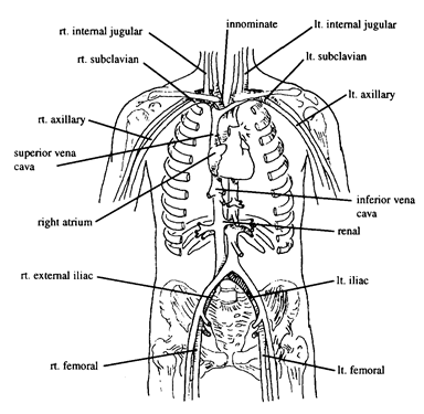
The vessels of the venous system parallel the arterial vessels. The blood returns to the heart by veins that lead to the superior and inferior vena cavae. These lead to the right atrium where the blood is moved through the ventricle and pulmonary artery to the lungs. It then returns to the left heart and enters the arterial system. |
|
|
Veins of the Head |
|
|
The veins of the brain drain into the venous sinuses of the brain. The saggital and transverse sinus veins drain the veins of the brain. The veins outside the skull and the sinuses inside the skull are connected by diploic veins. The venous blood from the head enters the external and internal jugular veins. These are bilateral veins. The external jugular vein is the largest vein in the neck. This vein drains the face and scalp, then enters into the subclavian vein. The internal jugular vein is deeper and runs alongside the carotid artery on both sides of the neck. It combines with the subclavian vein to become the innominate vein. As with the other veins, the innominate veins are bilateral. The innominate veins come together to form the superior vena cava. |
|
|
Vena Cava |
|
|
The vena cava consists of the superior and inferior vena cava. The vena cavae return venous blood to the right atrium. The superior vena cava drains the head, arms and upper body. The inferior vena cava drains the lower body and legs. |
|
|
Veins of the Upper Extremities |
|
|
The upper extremities have groups of deep and superficial veins. The subclavian vein drains the axillary vein from the arm. The brachial and basilic veins of the arms combine and drain into the axillary vein. The basilic vein is formed from the median basilic and ulnar veins. The axillary vein also combines with the cephalic vein before it becomes the subclavian. The cephalic vein runs in the arm up to the shoulder. The fingers are drained by digital veins that converge into the palmar arch. The metacarpal vein drains the palm of the hand. It also drains into the palmar arch. The arch joins with the radial vein that runs alongside the radial artery. Deep and superficial palmar veins connect the ulnar and radial veins. |
|
|
Large Veins |
|
|
There are two veins that receive blood from the stomach. These are the two gastroepiploic veins. The smaller of the two corresponds with the hepatic artery. The other corresponds with the gastric artery. They both drain into the inferior vena cava. Veins from the stomach, pancreas, intestines and the spleen drain blood rich in digested nutrients into the large portal vein. The portal vein branches to venules that empty into the liver. The capillaries of the liver then combine to form the hepatic vein that empties into the inferior vena cava. The splenic vein is formed from five or six branches that return blood from the spleen. The splenic vein and mesenteric veins combine to form the portal vein. The superior mesenteric vein drains blood from the small intestine and colon. The inferior mesenteric vein drains blood from the rectum and colon. These veins are parallel to their arteries. There are two renal veins, one from each of the kidneys. The renal veins empty into the inferior vena cava. Due to the filtering process of the kidneys, high volumes of blood are moved through these veins. |
|
|
Veins of the Lower Extremities |
|
|
The lower extremities are drained by a set of superficial veins and deep veins. The superficial veins, the greater and lesser saphenous veins drain the superficial lower leg, then join the deep veins. The deep veins are named after their matching arteries. The left common iliac and right common iliac veins combine to form the inferior vena cava. The iliac veins drain the lower extremities and pelvis The external iliac is an extension of the femoral vein. It joins with the internal iliac vein to form the common iliac vein. The femoral vein goes up the inner aspect of the thigh with the femoral artery to join with the greater saphenous vein to become the external iliac vein. The femoral vein drains most of the blood from the legs. |
|
|
The short, or lesser, saphenous vein begins at the outer arch on the top of the foot. It runs along the Achilles tendon to the popliteal vein. There are many branches of this vein receiving blood of the leg and foot. The long, or greater, saphenous, starts at the inner arch on the top of the foot. It moves up the inner thigh to join the femoral vein. These veins have many valves that help move the blood and prevent pooling. The greater saphenous vein is harvested to use for grafts during coronary bypass artery grafting. |
|
|
The popliteal vein receives blood from the anterior and posterior tibial veins and runs into the femoral vein. The anterior tibial vein runs between the tibia and fibula. This vein drains the knee, thigh and upper calf. It combines with the posterior tibial and popliteal veins. The posterior tibial vein is the vein before the lateral and medial plantar veins bifurcate. It runs alongside the tibial artery through the leg. |
|
|
Veins |
|
|
|
Уважаемый посетитель!
Чтобы распечатать файл, скачайте его (в формате Word).
Ссылка на скачивание - внизу страницы.