Cells, however, also transport certain materials in or out along gradients; this means that there is already more of a certain substance inside the cell than outside, for instance, but the cell needs even more. Movement along gradients (from region of lower concentration or pressure to region of higher concentration or pressure), like rolling a rock uphill, requires energy expenditure by the cell. Moving a substance along such a gradient involves the energy-cost by process of active transport. In many cell, active transport is accomplished by a set of proteins in the plasma membrane that act like pumps, pushing or pulling ions into and out of the cell.
One type is sodium – potassium pump. A red blood cell, for example, contains a high concentration of K+ but a low concentration of Na+, while the blood plasma outside the cell contains the reverse the special membrane proteins in the pump actively move K+ in and Na+ out along their concentration gradient by expending energy in the form of ATF. This pumping results in a proper osmotic balance, since water “follows” the Na+ out. Thus, so long as pumping continues, the cell does not shrink or burst.
RESTING AND ACTION POTENTIALS
1. Determination resting and action potentials
Certain
types of cell in living organisms, such as nerve and muscle fibers, are able to
respond and conduct electric signals. Nerve fibers consist of thin hollow tube
filled with aqueous electrolyte solutions containing ions of potassium (K+),
sodium (Na+), chlorine (Cl-). The fibers
are submerged into extracellular fluid. The walls of the tubes are a
semi-permeable membrane, which is also an electric insulator and allows ions to
migrate into and out of the fiber. Hence, membrane potential arises. The
membrane potential is a potential difference between internal and
external parts of a membrane ![]() . In its normal
state the membrane is permeable for potassium ions (K+). These ions
rapidly migrate from intercellular fluid to the extracellular one (Fig.1).
. In its normal
state the membrane is permeable for potassium ions (K+). These ions
rapidly migrate from intercellular fluid to the extracellular one (Fig.1).
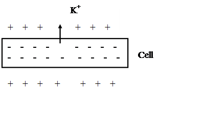 |
This process is called polarization. The inner part of a membrane acquires negative charge with respect to the external. This condition is called the membrane resting potential.
For dead cells the membrane potential, or Donane’s potential, is written as:
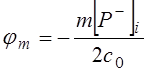 , (1)
, (1)
where [P-]I - is the concentration of albumen molecules inside of a membrane, m - the number of harge at the molecule surface, c0 - the ions concentration in extracellular fluid.
For living cells:
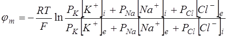 , (2)
, (2)
where R - the gas constant, T - the temperature, F - the Faraday’s number, PK,Na,Cl - the ions permeability; the i, e indexes denote concentration of ions inside and outside of a cell.
If an electric impulse of more than threshold value is applied to the nerve fiber, the membrane becomes more permeable for sodium ions (Na+) and this ions rapidly migrate from the extracellular fluid inside of the membrane, and the inside potential of the membrane becomes positive with respect to the external one. This process is called depolarization (Fig.2).
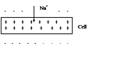 |
After a short period of time, which is called refractory period, the flow of sodium ions stops, the membrane becomes impermeable for ions and returns to its initial, or repolarization state. The electric impulse producing depolarization and the repolarization of cell is called action potential.
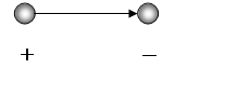
The configuration of two point charges, equal by value but opposite by polarity is called electric doublet or dipole (Fig.3).
 |
Basic positions of the theory:
·Heart is an electric doublet with integral vector of heart E. Vector E is geometrical sum of doublets of heart cells.
·The doublet is located in homogeneous conducting medium.
·The integral vector E changes its magnitude and direction in time. Origin of vector is stationary (is fixed), and its terminus is circumscribes a multiplex space-curve.
Practically, the potential difference of heart is measuring with the help of electrocardiography.
Electrocardiography is the method of investigation of electric activity heart. Method is based on the registration of time dependence of potential difference at the body surface. In this case the projection of multiplex space-curve of heart integral vector E is register in three electrocardiogram leads (in three plane):
· between the left and the right hands is the first electrocardiogram lead;
· between the right hand and the left leg is the se cond electrocardiogram lead;
· between the left hand and left leg is the third electrocardiogram lead.
However, for the receiving of full information about the heart functioning 12 leads are use.
Topic: Registration of heart acting potential with use of electrocardiograph “ЭКГ-01”
Devises: the electrocardiograph “ЭКГ-01”, electrodes, physiological/salt solution and napkins.
Work algorithm:
·Attach the electrodes accorging to elecrocardiogram leads,
·Register the heart acting potential in three elecrocardiogram leads,
·Calculate the cardiac bear rate (CBR) for each elecrocardiogram lead:
CBR=60v/R-R,
Where v is rate of movement of electrocardiogram tape, R-R is the distance between two R-tooth of the electrocardiogram tape.
·Make conclusion.
Уважаемый посетитель!
Чтобы распечатать файл, скачайте его (в формате Word).
Ссылка на скачивание - внизу страницы.